A big step in joint research
“The knee is the most exciting of all the joints in the human body,” says Bill Taylor, Professor of Movement Biomechanics at the Department of Health Sciences and Technology. “And it’s also the most complex. The knee joint is subject to tremendous acceleration and huge stresses, and it performs highly complicated movements.” Taylor discovered his passion for the knee some 20 years ago. Since then, his goal has been to understand every last detail of how this joint works. “First, we need to determine what kind of stresses act on the knee and what kind of movements it performs – only then will we be able to understand why the joint sometimes becomes stiff, or cartilage is depleted, or pain occurs,” he explains.
To learn more about how the knee works, Bill Taylor and his team are using an advanced form of videofluoroscopy – the imaging of skeletal structures within the human body by means of pulsed X-rays. Taylor combines this with an analysis of knee movement using skin markers, force plates and measurements of muscle activity. The Institute of Biomechanics at ETH Zurich first developed an automated moving fluoroscope over ten years ago in order to track the movement of the knee while walking and climbing stairs.
This arc-shaped X-ray device was mounted on a robot that followed the movements of the joint. As the test participants walked, the entire apparatus moved with them, thereby recording moving X-ray images of the knee. These two-dimensional images were then used to create three-dimensional reconstructions and anatomical models, including the muscles and ligaments of the knee joint. This showed the amount of stress exerted on individual ligaments during specific movements, which in turn is important for understanding how pain develops.
Unique measurement device
Although the original device provided much more accurate measurements than previous methods, it came with a number of drawbacks: images were in one plane only, and of a low resolution; likewise, on account of its design, only slow walking movements could be investigated. Taylor and his team therefore resolved to modify and enhance the original device. As Taylor explains, their vision was “to build a unique, state-of-the-art piece of equipment” that would set the gold standard, not only for basic research but also for clinical assessment of knee functionality. The goal was to provide answers to the following questions: Where best to begin with the rehabilitation of a damaged joint? When exactly does a knee joint need replacing? And which implant is best suited for a specific person?
The approval of a grant application to the Swiss National Science Foundation gave the green light for the development of the new dual-plane video fluoroscope. In addition to an innovative bi-planar imaging unit, the new device features electric motors that accelerate extremely quickly, thereby keeping the knee in the field of view of the imaging unit throughout the full cycle of stair climbing or other everyday activities. In addition, the device is able to track the movement of the subject by means of a single marker. This is attached to the subject’s knee but not to the fluoroscope, which means the subject is able to move freely.
Benefits of a new location
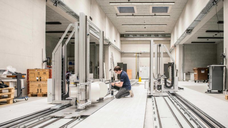
In autumn 2023, Professor Taylor and his team moved from the Hönggerberg campus to the new GLC building in the centre of Zurich. Transport and assembly of the new fluoroscope have posed major challenges, and both required lengthy planning. To ensure full functionality and compliance with all the safety regulations, the concrete foundations for the laboratory, which is located in the basement of the research building, had to be specially designed and constructed to accommodate the 22-metre-long test facility.
New ETH building GLC
With its new GLC building on the Gloriarank site, ETH Zurich has created an ultramodern lab and development facility in the Zurich City University District. Providing teaching, research and research translation in the fields of healthcare, medicine and medical technology, the cutting-edge facility was made possible by a generous donation from the Mäxi Foundation.
The proximity to clinics and other research institutions offers many advantages. “We work very closely with the Schulthess Clinic,” Taylor explains. “They helped us with a number of aspects, including the design of the new device.” Joint projects are also underway with Balgrist University Hospital, University Hospital Zurich and Kantonsspital Baden. The fluoroscope will be used for the first time at the new location to carry out a study of healthy test subjects. The aim here is to generate gold-standard data to enable a better understanding of the condition of intact knee joints. This will then serve as a benchmark for other studies, including an ongoing Innosuisse project that aims to examine study participants with artificial knee joints and deliver insights for the future development of implants.
Taylor, however, is already thinking several steps ahead. He would like to further reduce the already low radiation levels involved in this imaging process. This way, the technology could be used not only for peripheral joints such as the knee but also for more vulnerable parts of the body such as the shoulder or spine. Clinics are already beginning to show interest.
